Nature Communications, 2023
Computational pathology research
The design and capabilities of Slim are described in a peer-reviewed article on interoperable slide microscopy viewing and annotation for imaging data science.
- published in Nature Communications
- 2023
- standard annotation output
- TID 1500
The article Interoperable slide microscopy viewer and annotation tool for imaging data science and computational pathology (Gorman et al., Nature Communications 14:1572, 2023) explains why Slim stores everything in DICOM: images, the annotations experts draw on them, and the results AI models produce.
From annotation to model and back
Experts annotate regions in Slim, which stores them as DICOM Comprehensive 3D SR. Python libraries such as highdicom read those annotations to train models. The models write their output back as DICOM Segmentation, Parametric Map or Microscopy Bulk Simple Annotations, and Slim displays it next to the original slide.
Because every step uses the same standard, the loop works across institutions and archives without custom converters.
Open the data in Slim
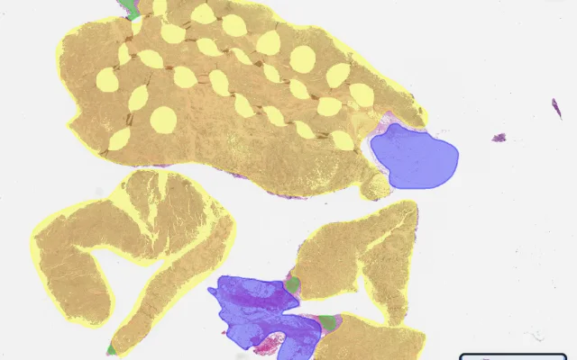
Comprehensive 3D SR
Expert region annotations stored as Comprehensive 3D SR(opens in the demo)
RMS-Mutation-PredictionOpen in Slim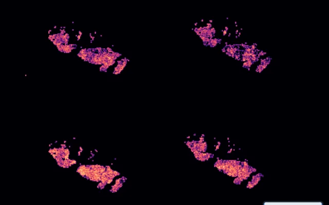
Parametric Map
Glioma aggressiveness score map, DICOM Parametric Map(opens in the demo)
TCGA-GBMOpen in Slim